
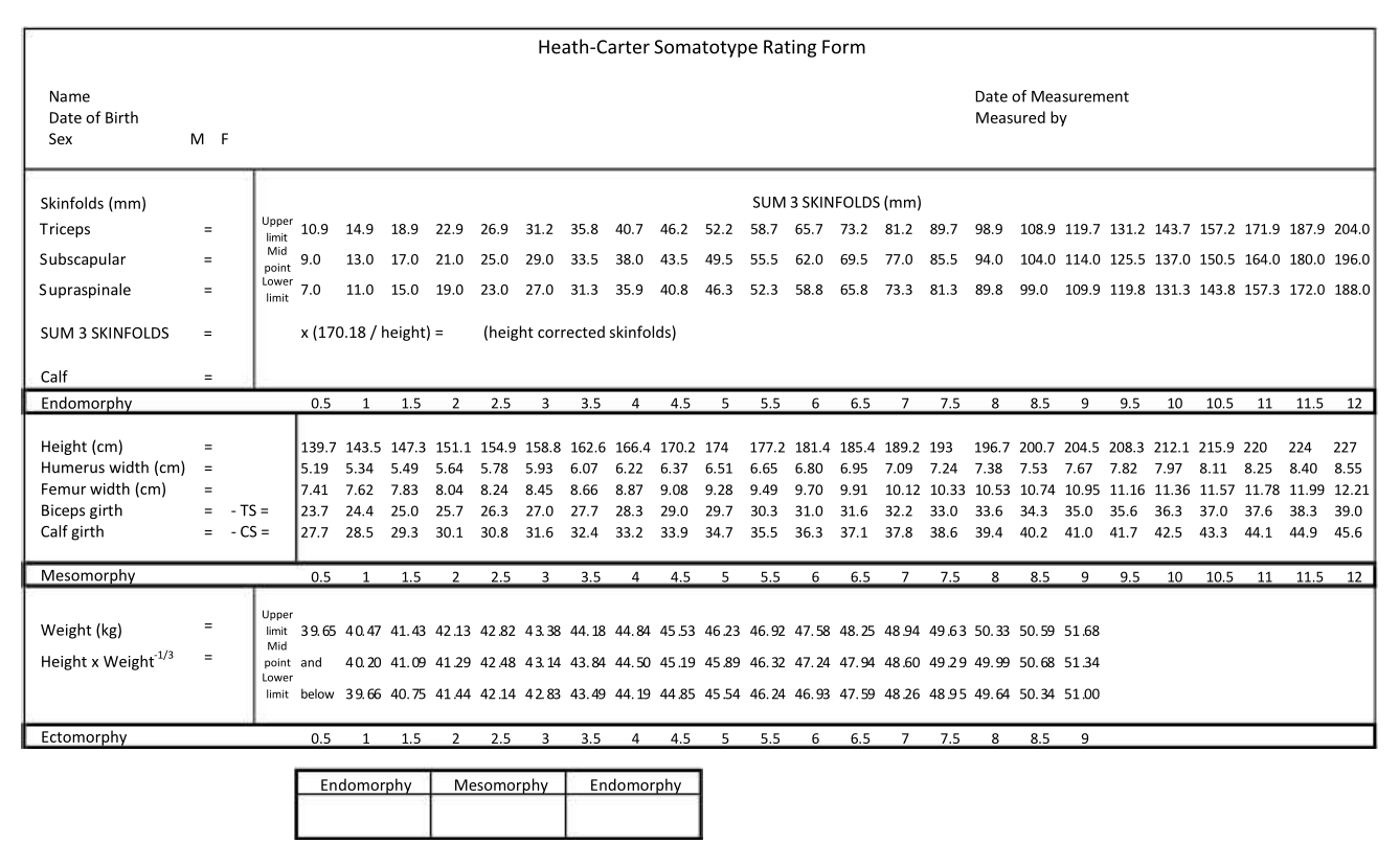
The Heath-Carter method of somatotyping is the most commonly used today. In each of the three categories someone is generally classified on a scale from 1 to 7 (though higher ratings are possible), though you cannot score highly on all three. The three numbers together give a somatotype number, with the endomorphy score first, then mesomorphy and finally ectomorphy (e.g. 1-5-2). The scores may also be plotted in a shield diagram or somatograph, representing the somatotype on a two dimensional scale.
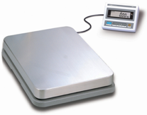
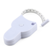
Anthropometric equipment includes a stadiometer or height scale and headboard, weighing scale, small sliding caliper, a flexible steel or fiberglass tape measure, and a skinfold caliper. The small sliding caliper is a modification of a standard anthropometric caliper or engineer’s vernier type caliper.
MEASUREMENTS FOR CALCULATING THE HEATH-CARTER ANTHROPOMETRIC SOMATOTYPE
Tools for Assessing Somatotype
Measurement Techniques
Ten anthropometric dimensions are needed to calculate the anthropometric somatotype: stretch stature, body mass, four skinfolds (triceps, subscapular, supraspinale, medial calf), two bone breadths (biepicondylar humerus and femur), and two limb girths (arm flexed and tensed, calf). The following descriptions are adapted from Carter and Heath (1990).
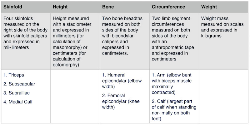
Stature (Height)
Taken against a height scale or stadiometer. Take height with the subject standing straight, against an upright wall or stadiometer, touching the wall with heels, buttocks and back. Orient the head in the Frankfort plane (the upper border of the ear opening and the lower border of the eye socket on a horizontal line), and the heels together. Instruct the subject to stretch upward and to take and hold a full breath. Lower the headboard until it firmly touches the vertex.
Body mass (Weight)
The subject, wearing minimal clothing, stands in the center of the scale platform. Record weight to the nearest tenth of a kilogram. A correction is made for clothing so that nude weight is used in subsequent calculations.
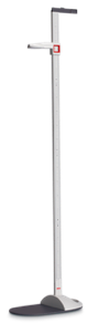
Skinfolds
Raise a fold of skin and subcutaneous tissue firmly between thumb and forefinger of the left hand and away from the underlying muscle at the marked site. Apply the edge of the plates on the caliper branches 1 cm below the fingers of the left hand and allow them to exert their full pressure before reading at 2 sec the thickness of the fold. Take all skinfolds on the right side of the body. The subject stands relaxed, except for the calf skinfold, which is taken with the subject seated.
Triceps Skinfold:
With the subject’s arm hanging loosely in the anatomical position, raise a fold at the back of the arm at a level halfway on a line connecting the acromion and the olecranon processes.
Subscapular Skinfold:
Raise the subscapular skinfold on a line from the inferior angle of the scapula in a direction that is obliquely downwards and laterally at 45 degrees.
Supraspinale Skinfold:
Raise the fold 5-7 cm (depending on the size of the subject) above the anterior superior iliac spine on a line to the anterior axillary border and on a diagonal line going downwards and medially at 45 degrees. (This skinfold was formerly called suprailiac, or anterior suprailiac. The name has been changed to distinguish it from other skinfolds called “suprailiac”, but taken at different locations.)
Medial calf Skinfold:
Raise a vertical skinfold on the medial side of the leg, at the level of the maximum girth of the calf.
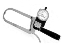
Biepicondylar Breadth of the Humerus
The width between the medial and lateral epicondyles of the humerus, with the shoulder and elbow flexed to 90 degrees. Apply the caliper at an angle approximately bisecting the angle of the elbow. Place firm pressure on the crossbars in order to compress the subcutaneous tissue.
Biepicondylar Breadth of the Femur
Seat the subject with knee bent at a right angle. Measure the greatest distance between the lateral and medial epicondyles of the femur with firm pressure on the crossbars in order to compress the subcutaneous tissue.
Upper Arm Girth (Elbow Flexed & Tensed)
The subject flexes the shoulder to 90 degrees and the elbow to 45 degrees, clenches the hand, and maximally contracts the elbow flexors and extensors. Take the measurement at the greatest girth of the arm.
Calf Girth
The subject stands with feet slightly apart. Place the tape around the calf and measure the maximum circumference.
Note:
Read stature and girths to the nearest mm, biepicondylar diameters to the nearest 0.5 mm, and skinfolds to the nearest 0.1 mm (Harpenden caliper) or 0.5 mm on other calipers.
Traditionally, for the anthropometric somatotype, the larger of the right and left breadths and girths have been used. When possible this should be done for the individual assessment.
Somatotype Categories
Somatotypes with similar relationships between the dominance of the components are grouped into categories named to reflect these relationships.
The frequencies of somatotypes within categories (or combined categories) can be used to describe the overall distribution of samples or for comparing them using a Chi-square analysis. The definitions of 13 categories are based on the areas of the 2-D somatochart (Carter and Heath, 1990).
- Central: no component differs by more than one unit from the other two.
- Balanced endomorph: endomorphy is dominant and mesomorphy and ectomorphy are equal (or do not differ by more than one-half unit).
- Mesomorphic endomorph: endomorphy is dominant and mesomorphy is greater than ectomorphy.
- Mesomorph-endomorph: endomorphy and mesomorphy are equal (or do not differ by more than one- half unit), and ectomorphy is smaller.
- Endomorphic mesomorph: mesomorphy is dominant and endomorphy is greater than ectomorphy.
- Balanced mesomorph: mesomorphy is dominant and endomorphy and ectomorphy are equal (or do not differ by more than one-half unit).
- Ectomorphic mesomorph: mesomorphy is dominant and ectomorphy is greater than endomorphy.
- Mesomorph-ectomorph: mesomorphy and ectomorphy are equal (or do not differ by more than one- half unit), and endomorphy is smaller.
- Mesomorphic ectomorph: ectomorphy is dominant and mesomorphy is greater than endomorphy.
- Balanced ectomorph: ectomorphy is dominant and endomorphy and mesomorphy are equal (or do not differ by more than one-half unit).
- Endomorphic ectomorph: ectomorphy is dominant and endomorphy is greater than mesomorphy.
- Endomorph-ectomorph: endomorphy and ectomorphy are equal (or do not differ by more than one-half unit), and mesomorphy is lower.
- Ectomorphic endomorph: endomorphy is dominant and ectomorphy is greater than mesomorphy.
The 13 categories can be simplified into four larger categories.
- Central: no component differs by more than one unit from the other two.
- Endomorph: endomorphy is dominant, mesomorphy and ectomorphy are more than one-half unit lower.
- Mesomorph: mesomorphy is dominant, endomorphy and ectomorphy are more than one-half unit lower.
- Ectomorph: ectomorphy is dominant, endomorphy and mesomorphy are more than one-half unit lower.