
Local muscle system (Local Stabilizers) includes the deep muscles that have their origin or insertion on the lumbar vertebrae. These muscles are closer to the center of rotation have shorter muscle lengths and are therefore ideal for controlling intersegmental motion. Key local stabilizing muscles include: Multifidus and Transversus Abdominus
Local System Symantec’s
- Deeper, smaller muscles
- Segmental control over 1 or 2 joint segments
- Control intersegmental motion
- Anticipatory activity
- Non-direction specific
- High resistance to fatigue
Muscles of the Local System
- Diaphragm
- Transversus abdominus
- Pelvic floor
- Multifidus
- Lower fibers of the internal obliques
- Deep fibers of psoas
- Deep fibers of erector spinae
- Medial fibers of quadratus lumborum
The Stabilization Mechanism
The local system (local musculature) refers to the core stabilization mechanism that consists of the deep muscles that attach to the vertebrae of the lumbar spine, pelvis and sacrum, allowing for segmental stabilization of the spine.
The local system has independent neurological control from that of the larger outer rectus abdominis, external oblique and anterior fibers of the internal obliques.
This would mean that many of the traditional gym exercises that are often used to condition the core are unable to condition the musculature of the inner unit effectively and may even lead to faulty recruitment patterns.
Failure to condition these muscles to optimal levels may and often do result in injury due to segmental instability, most often to the spine.
Specific isolation exercises are required accomplish automatic reflex control of the inner unit and to enhance sensory-motor control. Once control has been established, activation of the inner unit then can and must be programmed into all functional movement patterns.
Exercising the big muscles (prime movers), was not providing the correct strengthening for essential small muscles, such as the multifidus, transversus abdominis and pelvic floor. When working properly, these muscles provide the necessary increases in joint stiffness and stability to the spine, pelvis and rib cage to provide a stable platform for the big muscles. In a sense, as the big muscles (global system) became stronger and tighter the delicate balance between the local and global systems becomes disrupted.
When the local system is functioning correctly, joint injury is infrequent, even under extreme loads such as pushing a car, tackling an opponent in rugby or lifting large weights in the gym.
When it is not functioning correctly, activation of large prime movers will be no different than a large wind hitting a flag in the presence of loose guy wires running from vertebra to vertebra in the flag pole. As any system is only as strong as its weakest link.
Local system training provides essential joint stiffness and the stability needed to provide the large prime movers of the body with a working foundation. When global system or prime mover exercises are executed in absence of a functional inner unit, poor posture, unwanted aesthetic changes and musculoskeletal injury are inevitable. For optimal health and performance, the local system must not only be functional, but must be maintained with technically correct exercise protocol.
Core Stabilization Mechanisms
The Core or lumbo-pelvic-hip complex is stabilized during movement by three primary mechanisms; the thoracolumbar Stabilization, intra-abdominal pressure and the hydraulic amplifier mechanism.
- Thoracolumbar Fascia Stabilization
- Intra-Abdominal Pressure
- Hydraulic Amplifier Mechanism
These core stabilization mechanisms work synergistically to create stability throughout the core, in particular segmental stabilization through the lumbar spine itself.
Thoracolumbar Stabilization Mechanism
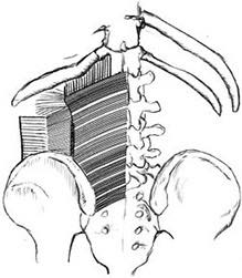
The thoracolumbar fascia (TLF) is an extensive fascial system in the back which consists primarily of three layers that include the posterior, anterior and middle. This essential system plays an important role in the functional stability of the lumbar spine.
Although the thoracolumbar fascia is non-contractile, it can be engaged dynamically because of the contractile tissue that attaches to it. These include the deep erector spinae, multifidus, transverse abdominis, internal oblique gluteus maximus, latissimus dorsi, and the quadratus lumborum.
The Thoracolumbar Fascia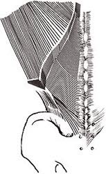
- Deep erector spinae
- Multifidus
- Transverse Abdominis
- Internal oblique
- Gluteus maximus
- Latissimus dorsi
- Quadratus lumborum
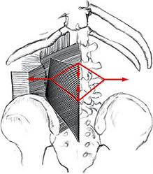
The TFL surrounds the erector spine and quadratus lumborum, thus providing support to these muscles when they contract. This can also be seen in the hydraulic amplifier mechanism as discussed below.
The transverse abdominis and the internal oblique are of particular importance for stabilization as they attach to the middle layer of the thoracolumbar fascia via the lateral raphe and create a traction and tension force on the thoracolumbar fascia when contracted, which enhances the regional intra-segmental stability in the lumbo-pelvic-hip complex.
This preferential contraction decreases translational and rotational stress at the lumbosacral junction.
Intra-Abdominal Pressure Mechanism
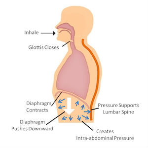
The intra-abdominal pressure mechanism (IAP) decreases compressive forces in the lumbo-pelvic-hip complex.
- Transverse Abdominis
- Posterior Fibers of Internal Oblique
- Diaphragm
- Multifidus
- Pelvic Floor Musculature
The muscles involved with the Intra-Abdominal pressure mechanism include the transversus abdominis, posterior fibres of the internal obliques, pelvic floor muscles, multifidus and the diaphragm.
It has been suggested that the Intra-Abdominal Pressure Mechanism contribute as much as 30 plus percent while others say as low as 10 percent to segmental stabilization of the lumbar spine.
As these abdominal muscles contract against the viscera, they push superiorly into the diaphragm and inferiorly into the pelvic floor. With this elevation of the diaphragm a decompression effect in L4 and L5 level of the lumbar spine is created. Along with the contraction of the pelvic floor musculature assists in providing intrinsic stabilization to the lumbo-pelvic-hip complex. This mechanism creates what is known as hoop tension.16
More recently, it has been shown that IAP does provide a stiffening effect on the lumbar spine, but that IAP is most effective at stabilizing the spine when applied in concert with co-activation of the erector spinae muscles.
Hydraulic Amplifier Mechanism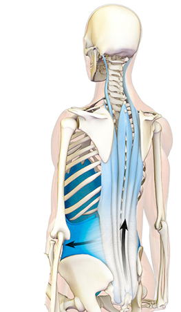
The hydraulic amplifier mechanism (HAM) has been shown to increases the strength of the back muscles through increased lumbar spine stiffness and its contribution to lumbar stabilization.
During flexion of the lumbar spine at approximately 45 degrees of flexion, the EMG activity of the erector spinae decreases, (this is referred to as flexion relaxation response) and “load-shifting” occurs to the non-contractile tissue (TFL) and the eccentrically contracting gluteals and hamstrings.
The TFL constrains the radial expansion of the three lumbar back muscles. Contraction of these muscles exerts a pushing force on the fascia creating potential energy that is then stored in the viscoelastic components of the non-contractile tissue (TFL) and in the parallel and series elastic elements of the eccentrically contracting gluteals and hamstrings.
ICP In-compartmental Pressure
Spring as much force can be generated as the contractional forces than be produced by the erector spinae.
During hip and trunk extension, the potential energy is converted into kinetic energy to assist the erector spinae in re-establishing an upright position.
This must be superimposed on an efficient thoracolumbar fascia mechanism. These stabilization mechanisms work interdependently to provide stabilization to the lumbo-pelvic-hip complex.