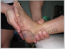
Extension of 1st MTP Joint
Motion occurs at the metatarsophalangeal joint in the sagittal plane around a coronal axis.
Normal Values:
0° to 70° (70°)
Testing Position:
Client is supine with knee extended with 0° of ankle dorsiflexion, plantarflexion, eversion, or inversion. The MTP joints of adjoining toes should be in neutral position. Flexion or extension of these MTP joints will stress across accessory tissues and may limit motion and obscure measurements. The IP (interphalangeal) joints are positioned in 0° of both flexion and extension.
Stabilization:
Prevent ankle dorsiflexion, inversion and eversion of the foot by stabilizing the metatarsal.
Goniometer Placement:
A: Medial MTP joint line.
SA: Medial midline of tarsal bone.
MA: Medial border proximal phalanx.
Assessment:
Passively extend 1st MTP Joint to the point of first resistance barrier and take measurement.
Possible Tight Structures:
Plantar surface musculotendinous tissues, including plantar fascia & flexor hallucis longus.
Recording the assessment
When recording the results make sure that any necessary notes are added to ensure intratester and intertester reliability.
![]()
Plantarflexion
Motion occurs at the talocrural joint in the sagittal plane about the coronal axis.
Normal Values:
0° to 50° (50°)
Testing Position:
Client is positioned supine with knee fully extended. Ankle is positioned in 00 of inversion and eversion at the subtalar joint. This position is known as “subtalar neutral.” Pinch the talar neck with thumb and index finger. Passively invert, then evert the foot until equal pressure is noted at the thumb and index finger. Hold that position, plantarflex the foot and take the measurement.
Stabilization:
Hold the tibia and fibula to prevent knee and hip rotation.
Goniometer Placement:
A: Center the goniometer fulcrum to the lateral aspect of the ankle.
SA: Lateral aspect of fibula.
MA: Midline of 5 metatarsal.
Assessment:
Passively plantarflex the foot to the first resistance bather without shifting from subtalar neutral.
Possible Tight Structures:
Anterior tibialis, peroneals, extensor digitorum longus, extensor digitorum brevis, extensor hallucis.
Recording the assessment
When recording the results make sure that any necessary notes are added to ensure intratester and intertester reliability.
Dorsiflexion (Gastrocnemius)
Motion occurs at the talocrural joint in the sagittal plane around the coronal axis.
Normal Values:
0° to 20° (20°)
Testing Position:
Client is positioned supine with knee fully extended. Ankle is positioned in 0° of inversion and eversion at the subtalar joint. This position is known as “subtalar neutral”. Pinch the talar neck with thumb and index finger. Passively invert, and then evert the foot until equal pressure is noted at the thumb and index finger. Hold that position to take the measurement.
Stabilization:
Hold the tibia and fibula to prevent knee and hip rotation.
Goniometer Placement:
A: Center the goniometer fulcrum to the lateral aspect of the ankle.
SA: Lateral aspect of fibula.
MA: Midline of 5th metatarsal.
Assessment:
Passively dorsiflex the foot to the point of first resistance without shifting out of “subtalar neutral.” Hold and take measurement.
Possible Tight Structures:
Gastrocnemius, soleus, posterior tibialis
Recording the assessment
When recording the results make sure that any necessary notes are added to ensure intratester and intertester reliability.
![]()
Dorsiflexion (Soleus)
Motion occurs at the talocrural joint in the sagittal plane around the coronal axis.
Normal Values:
0° to 20° (20°)
Testing Position:
Client is positioned supine with knee flexed at 30°. Ankle is positioned in 0° of inversion and eversion at the subtalar joint. This position is known as “subtalar neutral”. Pinch the talar neck with thumb and index finger. Passively invert, and then evert the foot until equal pressure is noted at the thumb and index finger. Hold that position to take the measurement.
Stabilization:
Hold the tibia and fibula to prevent knee and hip rotation.
Goniometer Placement:
A: Center the goniometer fulcrum to the lateral aspect of the ankle.
SA: Lateral aspect of fibula.
MA: Midline of 5th metatarsal.
Assessment:
Passively dorsiflex the foot to the point of first resistance without shifting out of “subtalar neutral.” Hold and take measurement.
Possible Tight Structures:
Soleus and posterior tibialis muscle.
Recording the assessment
When recording the results make sure that any necessary notes are added to ensure intratester and intertester reliability.