
Physiology of the Eye
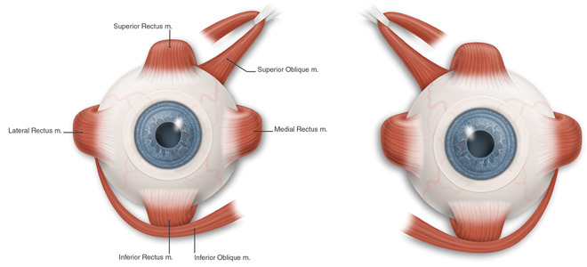
Each eye is moved by six muscles, controlled by three cranial nerves (III, IV and VI).
When you look straight ahead, the 4 rectus muscles of each eye do most of the work of aligning your eyes.
The oblique muscles come more into play on turning or tilting your head.
Each eye sends its own picture to the brain. The brain compares the images, and sends signals to the nerves controlling the extraocular muscles to coordinate the eyes and prevent double vision.
The hook in the superior oblique muscle is where it passes through a little pulley at the upper, inner margin of the bony orbit around your eye.
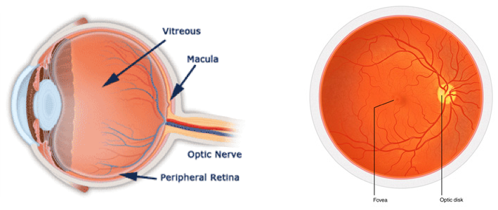
The retina lines the inside of the back of the eye.
It is composed of the macula and the peripheral retina.
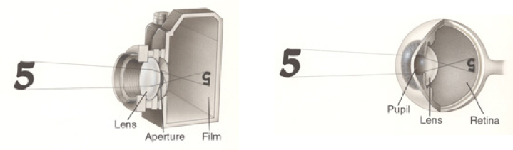
The retina can be compared to the film in a camera (or the light sensor in a digital camera).
Light coming into the eye passes through the cornea and lens (function like a camera lens) and focuses on the retina (which functions like the film or image sensor).
The image on the retina is upside down, just like a camera, but when the brain processes the image it turns right side up.
The fovea is where the finest detail vision is perceived; both the fovea and the surrounding macula perceive color.
The retinal periphery have lower visual acuity and it functions better than the macula at night, the peripheral retina has poor color vision.
The optic nerve enters the back of the eye. It carries the vision message from the retina back to the brain.
When a specialist looks into the back of the eye, the visible portion of then optic nerve is called either the optic disk or the optic nerve head.
The central part of the retina is called the macula, and its very center is the fovea.
Optic Flow
Optic flow is defined as the change of structured light in the image, e.g. on the retina, due to a relative motion between the eyeball and the scene.
Optic flow can also induce postural sway in the absence of physical movement perturbations (Lee and Aronson 1974; Lestienne et al. 1977)
What is Visuomotor Function
Visuomotor function is the integration between visual perception and motor skills.
Visual-motor function involves the ability to coordinate vision with the movements of the body.
Due to the body, head, eyes, and limbs being constantly in motion, every time an individual wants to carry out an action, calculations and decisions about orientation, motion, and location need to be made.
The parietal cortex is the part of the brain that is responsible for processing and integrating somatosensory, visual, and auditory information and plays an important role in producing planned movements.
The cerebellum, brainstem, and frontal lobe are also involved in visuomotor function.
The inner ear connection
The inner ear does not play as major role in balance as once thought.
The inner ear is dependent upon the eyes and actually stabilizes the eye in movement.
The inner ear is a fail safe when other systems have failed.
The 4 Basic Visual Skills
- Eye Movements
- Visual Clarity
- Depth Judgment (Perception)
- Peripheral Awareness