
Postural Reflexes are automatic movements that control the equilibration we require once upright and moving and having to combat the effects of gravity. They maintain posture, balance and fluidity of movement, replacing the Primitive Reflexes in a sequential manner, as those Primitive Reflexes inhibit. The first to emerge is the head righting reflex on a vertical plane. At birth the baby has no head control, they then develop conscious control to lift their head; lowering the head at that stage is more difficult. Eventually this movement is automated so that the head remains centrally aligned on top of the body with the crown uppermost. Tip a new born upside down and their head will just hang, tip a 12 week old baby and the head will extend backwards so that the crown is uppermost. Over time this ability to maintain head position develops on all planes. Another Postural Reflex is automatic flexion of the knee whenever the hip is rotated; this has obvious importance in walking and balance.
These reflexes are controlled by interplay between the sensory vestibular system; in the inner ear, the cerebellum and motor nerves controlling the muscles, as are the Primitive Reflexes but the area of control is higher up the brain stem within the mid brain. The development of the fibres creating stronger connections upwards is the result of normal neuro-development. Development between the lower and higher regions of the brain stem is never all or nothing; so that the majority of adults have deficits in this area and the reason that most of us, regardless of years of training, would not become top athletes. Children with Neuro-Developmental Delay (NDD) have less development in this area than is normal; the amount varying with the extent of delay and hence difficulty. There is inevitably a relationship between the number and strength of the retained Primitive Reflexes and that of the Postural Reflexes. This is the reason why so many children with learning difficulties have co-ordination and balance problems. Many parents will tell me that their child, though having reading difficulties, is exceptionally well co-ordinated. These are often children with less profound learning difficulty; children who have done a lot of sport and have well developed specific Postural Reflexes; or children who have developed all Postural Reflexes, but the connections are not as strong as they should be. These are the children that when put under pressure cannot maintain physical, emotional or academic competence. They fall to pieces in exams, cannot support physical prowess under slow or extreme circumstances.
The degree of learning difficulty is usually in direct relationship with the ability to automatically control the eyes. Eyes position is maintained by 6 paired muscles, attached to the outside of each eye. To clearly perceive an image the two eyes must be in alignment with the image and with each other. This is achieved by developing a dominant eye, with the other, as slave, lining up in response to its master. If this is not achieved then the image fails to be correctly fused and the individual will see double; for brief periods it is possible to consciously compensate, so that the image will fuse, fragment, fuse and fragment. Reading and copying are very difficult given this situation. The difficulty will be directly related to the degree of alignment error. The eye position in the orbit also needs to change according to whether the eye is looking at something far away, or near to; accommodation of the eye for distance is not entirely within the realms of the internal lens of the eye. Individuals with retained Primitive Reflexes frequently cannot manage this adjustment; with neither eye accommodating, or with only one. The degree of difficulty will depend on the degree and type of error; from poor definition to double image. For effective eye function we must have inhibited the Primitive Reflexes and developed effective Postural Reflex control from strong neural links between the vestibular system and the external muscles of the eye.
The relationship between the Primitive and Postural Reflexes can best be illustrated by the relationship between the foundations and upper structure of a house. If the foundations are weak then the house will begin to slip, the degree of slippage dependent upon the weakness in the foundations and the pressure from above on the structure of the house. This explains why the degree of difficulty experienced becomes greater the greater the pressure. The greater the importance of competence, and the more that competence is being measured, the greater the stress. All sensory input, other than taste and smell, is directed to the higher regions of the brain through the brain stem. The underdeveloped nerve pathways within the brain stem can cope more effectively the less the number of impulses transmitted. This is also why multi-sensory teaching, though very beneficial for the child with learning difficulties, must not be simultaneous; otherwise the child experiences the very real likelihood of sensory overload with the brain closing down because it cannot process or respond to any.
The Postural Reflexes:
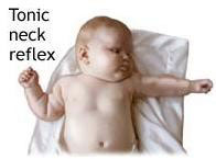
- The Transformed Tonic Neck Reflex
- The Amphibian Reflex
- Segmental Rolling Reflexes
- Occulo-Head Righting Reflex
- Labyrinthine-Head Righting Reflex
Development of postural reflexes begins in the womb with the development of primal reflexes. Primal reflexes are developmental and should not be retained but rather are processed into what is know as postural reflexes.
The postural reflexes support control of balance, posture and movement in a gravity based environment.
Postural Reflexes are automatic movements that control the equilibration we require once upright and moving and having to combat the effects of gravity.
They maintain posture, balance and fluidity of movement, replacing or controlling the Primitive Reflexes in a sequential manner, as those Primitive Reflexes are inhibited or controlled.
- The Plantar Reflex
- Asymmetrical Tonic Neck Reflex
- The Tonic Labyrinthine Reflex (TLR) Forwards
- The Tonic Labyrinthine Reflex (TLR) Backwards
Plantar reflex is a grasping reflex, balance and mobility, enhances myelination CNS.
Rooting reflex.
Postural reflexes are transformed primitive reflexes and executed by the cortex.
Bridging Postural Reflexes and should not be retained.
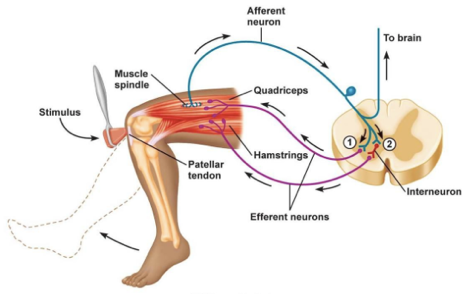
Reflex Arc Of Postural Reflex
Afferent Pathway comes from the eyes, the vestibular apparatus and the proprioceptors.
Integrating Centers are formed by neuronal network in the brain stem and spinal cord.
Efferent Pathway α-motor neurons supplying the various skeletal muscles i.e. the effector organ
Reflex Arch is the nerve pathway involved in a reflex action, including at its simplest a sensory nerve and a motor nerve with a synapse between.
The reflex arc is the basic unit of integrated reflex activity.
The reflex arc has 5 components:
- Receptor – muscle spindle ( DTR)
- Afferent – 1a , II fibers
- Centre – spinal cord
- Efferent – motor nerve
- Effector – extrafusal fibers.
Types Of Postural Reflexes
There are two types of postural reflexes:
- Static Reflexes
- Statokinetic Reflexes
Both these types of postural reflexes are integrated at various levels in the CNS from the spinal cord to cerebral cortex and are affected largely by pyramidal pathways.
Static Reflexes
Static reflexes are involved in adjustments to displacements produced by gravity. There are three types of static reflexes:
- Local static reflexes
- Segmental static reflexes
- General static reflexes
Local Static Reflexes
They exert their effect on the same limb from which the stimulus was initiated
The centre of local static reflex are located in spinal cord
Some important local static reflexes include:
- Stretch reflex
- Positive supporting reaction
- Negative supporting reaction
Stretch Reflex
This is the most important local static reflex which controls the tone in those extensor muscles which keep the body upright (antigravity muscles)
Positive Supporting Reaction
It is characterized by simultaneous reflex contraction of both extensors and flexors of a limb It plays an important role of steadying the ankle joint in standing position
Negative Supporting Reaction
It refers to disappearance of positive supporting reaction. It is initiated by stretch of the extensor muscles
Segmental Static Reflexes
It is characterized by a bilateral reflex response when stimulus is applied to one limb
The best example of segmental static reflex is crossed extensor reflex response component of withdrawal reflex
The centre of this reflex is in spinal cord
General Static Reflexes
It is characterized by a generalized effect from many muscle groups in the body in response to a stimulus that arises at one side of the body.
General static reflexes can be divided into thee groups:
- Attitudinal reflexes
- Long loop stretch reflexes
- Righting reflexes
Attitudinal Reflexes
These reflexes are initiated when the attitude of the body is changed i.e. while standing on an inclined plane.
These reflexes are of two types:
- Tonic labyrinthine reflex
- Tonic neck reflex
Tonic Labyrinthine Reflex
These reflexes are produced in response to alternation in position of head relative to horizontal plane e.g. while standing on an inclined plane.
- Pathway of reflex arc-
- Stimulus- is gravity
- Receptors- otolith organ (utricle and saccule of labyrinth)
- Afferents- from receptors travel along the vestibular nerve
- Centre- vestibular and reticular nuclei present in spinal cord
- Efferents- vestibulospinal and reticulospinal tracts which end on α-motor neurons of spinal cord
- Reflex reponse- contraction of extensor muscles of limb
Tonic Neck Reflex
These reflexes are produced in response to alternation in the position of head relative to the body.
- Pathway of reflex arc-
- Stimulus- stretch of neck muscles
- Receptors- pacinian corpuscles in the ligaments of the cervical joint and muscle spindle of neck muscles
- Centre- in the medulla oblongata
- Reflex response- depends on the position of the head in relation to the body:
- Turning down of head causes flexion of the forelimb and extension of the hind limb.
- Turning up of head causes reverse of the above.
- Turning the head sideways produces flexion of.
Long Loop Stretch Reflex
Long Loop Stretch Reflex is also called functional stretch reflex.
These are polysynaptic reflexes with the centre of reflex arc in cerebral cortex. These reflexes are continuously active in erect posture. They bring about continuous correction of the sways that occur from moment to moment during standing.
Righting Reflexes
Righting reflexes help to correct the position of the body when it goes off balance and falls down
These reflexes consists of a chain of reactions following one another in an orderly sequence
For example, if an animal is laid on it’s side or back, head rights itself followed by body and animal finally resumes upright posture.
The sequence of righting reflexes will be as follows:
Head righting reflex or Labyrinthine righting reflex
It is initiated when animal’s head is in lateral position.
Impulses arising from the saccules reflexly stimulate the appropriate muscles to bring head back to upright position
Body righting reflex
When the animal lies on the ground, the side in contact with the ground is constantly stimulated while the other side is not.
This differential stimulation of the deep structures in the body wall reflexly rights the head
Neck righting reflex
The head is righted by above two reflexes but the body still remains in lateral position
This leads to twisting of neck and this brings thorax and lumbar region successively into upright posture.
If the righting of head is prevented , impulses from the body surface may cause righting of the body directly (Body on body righting reflex).
Righting Reflexes
– Limbs righting reflex
– Impulses arising from the limb muscles leads to attainment of appropriate posture of limbs
– Optical righting reflexes
– Optical impulses also cause righting of the head in animals with intact visual cortex
– Centers Of Righting Reflexes
Chief center for all righting reflexes, except the optical righting reflex is red nucleus lying in the mid brain.
The center for optical righting reflex is in the visual cortex.
Statokinetic Reflexes
Statokinetic reflexes include:
– Vestibular placing reaction
– Visual placing reaction
– Hopping reaction
Vestibular placing reaction
– Stimulus: linear acceleration
– Receptors: in utricle and saccule
– Center: cerebral cortex
– Response: as soon as foot comes in contact of any firm surface, it is reflexly placed on surface in position to support the body
Visual placing reaction
– Stimulus: visual cues
– Receptors: eyes
– Center: cerebral cortex
– Response: as in above
Hopping reaction
– Stimulus: lateral displacement while standing
– Receptors: muscle spindle
– Center: cerebral cortex
– Response: hopping movement that keeps the limb in position to support the body when standing animal is pushed laterally