When a chain reaction evolves in which some muscles shorten and others weaken, predictable patterns involving imbalances develop.
Vladimir Janda, a Czech researcher, labelled these upper and lower ‘crossed’ syndromes.
The more common postural distortion patterns include:
- Lower cross syndrome
- Upper cross syndrome
- Sway Back Posture
- Military Type Posture
- Flat Back Posture
- Pronation distortion syndrome
- Head Forward Posture
- Scoliosis
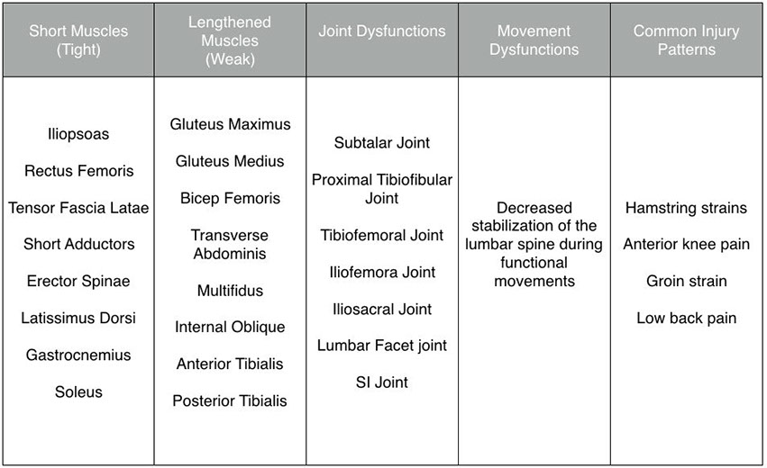
Lower Cross Syndrome
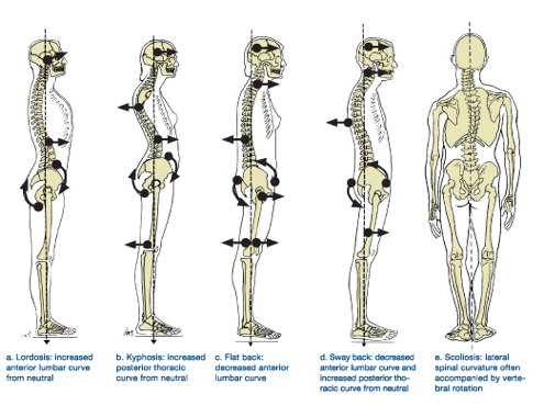
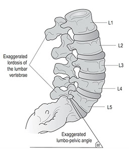
The lower cross syndrome is characterized by an increased lumbar lordosis and an anterior pelvic tilt.
The common muscular imbalances seen with the lower cross syndrome include short and tight hip flexors and lumbar erectors (erector spinae) crossed with lengthened and inhibited gluteus maximus and lumbo-pelvic-hip stabilizing mechanism (transverse abdominis, internal oblique, multifidus, pelvic floor muscles and diaphragm).
Also commonly involved are the piriformis muscles which in 20% of individuals are penetrated by the sciatic nerve so that piriformis syndrome can produce direct sciatic pressure and pain. Arterial involvement of piriformis shortness produces ischaemia of the lower extremity, and through a relative fixation of the sacrum, sacroiliac dysfunction and pain in the hip.
Potential Sources of Pain
Stress to the anterior longitudinal ligament
Narrowing of the posterior disk space and narrowing of the intervertebral foramen. This may compress the dura and blood vessels of the related nerve root or the nerve root itself, especially if there are degenerative changes in the vertebra or disk.
Approximation of the articular facets. The facets may become weight bearing, which may cause synovial irritation and joint inflammation.
Potential Muscle Impairments
Decreased flexibility in the hip flexor muscleiopsoas, tensor fasciae latae, rectus femoris, lumbar extensor muscles (erector spinae)
Stretched and weak abdominal muscles (rectus abdominis, internal and external obliques, and transverse abdominis)
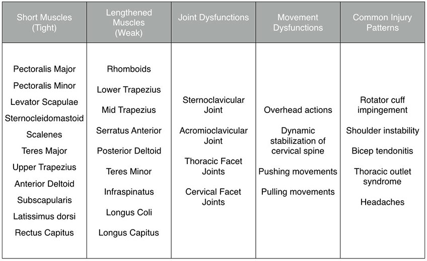
Upper Cross Syndrome (Kyphosis)
The upper cross syndrome or kyphosis is characterized by rounded shoulders and a forward head posture. This pattern is common in individuals who sit a lot or who develop pattern overload from uni-deminsional training protocols.
The common muscular imbalances seen with the upper cross syndrome include short and tight latissimus dorsi, pectoralis major and minor, upper trapezius, levator scapulae, and sternocleidomastoid (SCM) crossed with lengthened, weak stabilizers of the scapula (serratus anterior, rhomboids, middle and lower trapezius) and deep neck flexors.
The result of these changes is greater cervical segment strain plus referred pain to the chest, shoulders and arms. Pain mimicking angina may be noted plus a decline in respiratory efficiency.
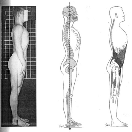
Flat Back Posture
The Flat Back Posture is characterized by a decrease lumbar lordosis and a posterior pelvic tilt. This is usually coupled with a flat thoracic spine.
The common muscular imbalances seen with the Flat Back Posture include short and tight hamstring musculature and lumbar erectors (erector spinae) crossed with lengthened and inhibited gluteus maximus and lumbo-pelvic-hip stabilizing mechanism (transverse abdominis, internal oblique, multifidus, pelvic floor muscles and diaphragm).
Also commonly involved are the piriformis muscles which in 20% of individuals are penetrated by the sciatic nerve so that piriformis syndrome can produce direct sciatic pressure and pain.
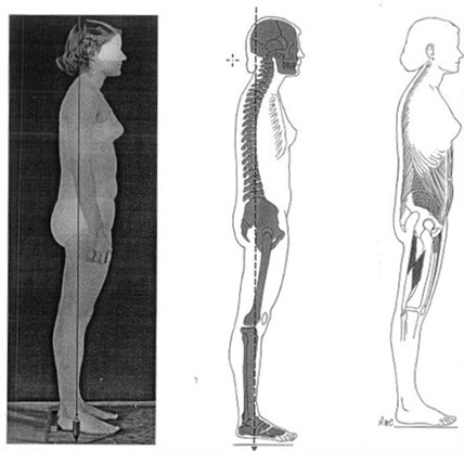
Sway Back Posture
With the Sway Back Posture the hamstrings and lower abdominals are short and tight, while the lumbar erectors, rectus femoris and iliopsoas are long and may be weak.
Note: The hips sway forwards of the line of good posture – hence the name.
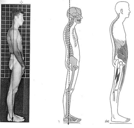
Military Type Posture
The Military Type Posture is characterized by hyperextension in the knees, the pelvis is tilted interiorly, and there is an increased lumbar lordosis.
The effect this posture has on the muscular system is predictable; the rectus abdominis muscle is elongated, the hip flexor muscles and low back muscles have a tendency toward shortening, and the hamstrings are lengthened past their ideal resting length.
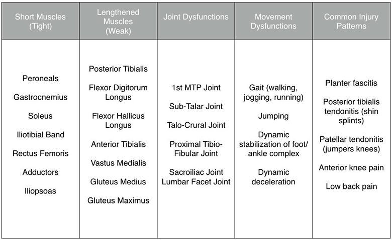
Pronation Distortion Syndrome
The lower extremity postural distortion is characterized by excessive foot pronation (flat feet), knee flexion coupled with internal rotation and valgus (knock-kneed) during functional movements.
The common muscular imbalances seen with the lower-extremity postural distortion include short and tight gastrocnemius, soleus, peroneals, adductors, iliotibial band (IT band) and hip flexors crossed with lengthened, weak anterior and posterior tibialis, vastus medialis (VMO), gluteus medius and hip external rotators.
When one foot exhibiting greater pronation than the other this can put an axial torque on the spine and can facilitate a number of challenging orthopedic and postural problems.
To assess, simply put the patient in sub-talar neutral, leaving them standing that way and reassess iliac crest height, shoulder height and rotation of the extremities and cranium. You may find that there are torque syndromes that must be corrected via an orthotic device.
Note: Flat Feet are also commonly related to nutritional deficiency in the parents.